
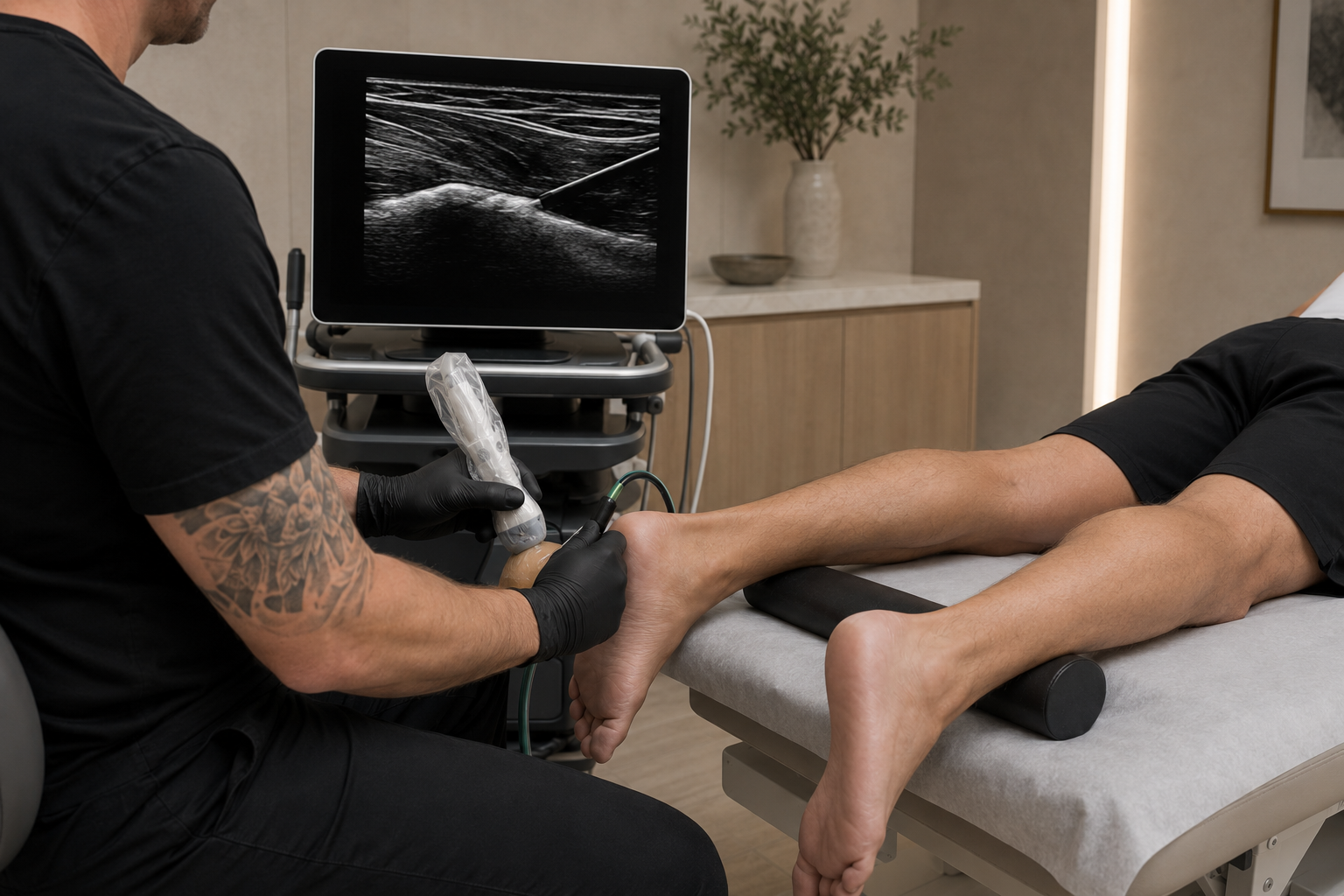
TENEX AND TENOTOMY
TENEX and Needle Tenotomy
Minimally Invasive Treatment for Chronic Tendon Pain and Degenerative Tendon Injuries
Chronic tendon pain can persist even after rest, physical therapy, medication, bracing, activity modification, or injections. In many of these cases, the problem is not ongoing short-term inflammation. Instead, the tendon may contain chronically damaged, disorganized, or degenerative tissue that is no longer progressing through a normal healing response.
TENEX and ultrasound-guided needle tenotomy are minimally invasive procedures designed to treat this type of chronic tendon dysfunction.
Both procedures target abnormal tendon tissue under direct musculoskeletal ultrasound guidance. The goal is to stimulate a new, organized healing response and improve the tendon’s ability to tolerate movement, exercise, work, and athletic activity.
TENEX uses ultrasonic energy with irrigation and aspiration to break down and remove selected damaged tissue. Needle tenotomy uses a standard medical needle to repeatedly stimulate the abnormal portion of the tendon without removing it.
These procedures can provide an intermediate treatment option between conservative care and traditional open surgery.
What Is Tendinopathy?
Tendinopathy is a chronic condition affecting a tendon—the strong connective tissue that attaches muscle to bone.
Although chronic tendon pain is often called tendinitis, many long-standing tendon conditions are not primarily inflammatory. The tissue may instead demonstrate:
Disorganized collagen
Tendon thickening
Partial fiber disruption
Reduced blood flow
Abnormal blood-vessel formation
Calcification
Scar tissue
Loss of normal tendon stiffness
Degenerative tissue changes
Reduced ability to tolerate mechanical loading
This chronic degeneration is sometimes described as tendinosis.
Pain may improve temporarily with rest or medication, but symptoms frequently return when the tendon is loaded again. This can create a cycle of pain, reduced activity, weakness, and repeated irritation.
TENEX and needle tenotomy are designed to interrupt this cycle by targeting the abnormal tendon tissue and stimulating a renewed healing response.
What Is Percutaneous Tenotomy?
The term percutaneous means that the procedure is performed through the skin rather than through a large surgical incision.
A tenotomy is a procedure involving a tendon. In minimally invasive sports medicine, percutaneous tenotomy generally refers to using a needle or small specialized instrument to stimulate, fragment, or remove chronically damaged tendon tissue.
The procedure is performed under musculoskeletal ultrasound guidance so the provider can:
Identify the abnormal tendon tissue
Visualize the treatment instrument in real time
Avoid nearby nerves and blood vessels
Target the damaged tissue precisely
Limit disruption of healthier tendon fibers
Evaluate the surrounding anatomy
Percutaneous needle tenotomy and TENEX ultrasonic tenotomy use different instruments, but both are based on the principle that chronic degenerative tendon tissue may benefit from controlled mechanical treatment.
What Is the TENEX Procedure?
TENEX is a minimally invasive form of percutaneous ultrasonic tenotomy.
The procedure uses a specialized microtip that delivers ultrasonic energy into the diseased portion of a tendon or fascia. The ultrasonic energy fragments and emulsifies selected abnormal soft tissue, while irrigation and aspiration help remove the treated tissue.
The FDA-cleared TENEX Health TX System is classified as an ultrasonic surgical aspirator and is indicated for procedures in which fragmentation, emulsification, and aspiration of soft tissue are desired, including orthopedic procedures.
The procedure is performed through a small skin opening and generally does not require the larger incision, sutures, or extensive tissue exposure associated with open tendon surgery.
How TENEX Works
TENEX treatment involves several coordinated steps.
Identifying the Damaged Tissue
Musculoskeletal ultrasound is used to locate areas of tendon thickening, degeneration, tearing, calcification, or abnormal tissue architecture.
The provider can compare healthy and unhealthy areas of the tendon and identify the safest treatment path.
Local Anesthesia
The skin and surrounding treatment area are numbed with local anesthetic. General anesthesia is typically unnecessary.
Introducing the Microtip
A small opening is created in the skin, and the TENEX microtip is advanced toward the abnormal tissue under direct ultrasound guidance.
Ultrasonic Tissue Treatment
The microtip delivers ultrasonic energy that fragments and emulsifies selected abnormal soft tissue.
Irrigation and Aspiration
The system introduces fluid while simultaneously removing fragmented tissue. This allows the provider to selectively debride the targeted area through a very small access point.
Closing and Protecting the Area
The microtip is removed, and the small opening is covered with a dressing. Sutures are often unnecessary, although the exact closure method depends on the treatment area and procedure performed.
What Is Ultrasound-Guided Needle Tenotomy?
Needle tenotomy—also called percutaneous needle tenotomy, tendon fenestration, or dry needling of the tendon—is performed using a standard medical needle.
Under ultrasound guidance, the needle is passed through the abnormal portion of the tendon multiple times in a controlled pattern.
This creates small channels within the degenerative tissue and converts a chronic, biologically inactive condition into a controlled acute healing response. The intended result is renewed bleeding, cellular recruitment, inflammatory signaling, collagen turnover, and tissue remodeling.
Unlike TENEX, conventional needle tenotomy does not use ultrasonic energy or aspiration to remove tissue.
How Needle Tenotomy Works
During ultrasound-guided needle tenotomy:
The injured tendon is evaluated with musculoskeletal ultrasound.
The skin and treatment area are sterilized.
Local anesthetic is administered.
A needle is advanced into the abnormal portion of the tendon.
The needle is passed through the damaged tissue in multiple directions.
Calcific, scarred, or degenerative areas may be mechanically disrupted.
The needle is removed, and a small dressing is applied.
The controlled needle passes stimulate localized bleeding and biological signaling within the abnormal tendon.
Needle tenotomy may be performed alone or combined with another regenerative medicine treatment, such as platelet-rich plasma or plasma prolotherapy.
TENEX Versus Needle Tenotomy
Both procedures are designed to treat chronic tendon or fascial disease, but they do so differently.
TENEX Ultrasonic Tenotomy
TENEX:
Uses a specialized ultrasonic microtip
Fragments selected abnormal tissue
Uses irrigation and aspiration
Physically removes a portion of the treated tissue
Requires a small skin opening
Is generally used for more established or focal degenerative tissue
May be useful when ultrasound demonstrates scarred, thickened, calcific, or clearly diseased tissue
Percutaneous Needle Tenotomy
Needle tenotomy:
Uses a standard medical needle
Creates multiple controlled passes through the tendon
Stimulates bleeding and a renewed healing response
Does not aspirate or remove tissue
Typically requires only a needle puncture
May be appropriate for less extensive tendinopathy
Can be combined with PRP or another orthobiologic treatment
Neither procedure is automatically superior for every patient.
The correct treatment depends on the location, tissue appearance, severity of degeneration, duration of symptoms, previous treatments, functional demands, and rehabilitation goals.
Conditions Commonly Treated With TENEX or Needle Tenotomy
These procedures may be considered for chronic tendon or fascial conditions that have not improved adequately with conservative care.
Elbow
Lateral epicondylitis, or tennis elbow
Medial epicondylitis, or golfer’s elbow
Common extensor tendinopathy
Common flexor tendinopathy
Distal triceps tendinopathy
Selected distal biceps tendon conditions
Shoulder
Rotator cuff tendinopathy
Calcific rotator cuff tendinopathy
Selected partial tendon injuries
Chronic biceps tendinopathy
Not every rotator cuff tear is appropriate for percutaneous tenotomy. Full-thickness tears, major retraction, muscle atrophy, instability, or advanced joint disease may require a surgical consultation.
Hip and Pelvis
Gluteus medius tendinopathy
Gluteus minimus tendinopathy
Greater trochanteric pain syndrome
Proximal hamstring tendinopathy
Selected adductor tendon injuries
Iliotibial band-related pathology
Tensor fascia lata tendinopathy
Knee
Patellar tendinopathy
Quadriceps tendinopathy
Jumper’s knee
Selected iliotibial band conditions
Chronic tendon attachment pain
Foot and Ankle
Plantar fasciopathy
Chronic plantar heel pain
Achilles tendinopathy
Insertional Achilles tendinopathy
Peroneal tendinopathy
Posterior tibial tendinopathy
Other Conditions
Chronic tendon scarring
Calcific tendon deposits
Recalcitrant tendon pain
Chronic myotendinous injuries
Selected postoperative scar or tendon abnormalities
Evidence is strongest for certain conditions, particularly chronic elbow tendinopathy and plantar fascia pain. Research involving other tendon sites is promising but remains more limited and heterogeneous.
When TENEX May Be Recommended
TENEX may be considered when:
Symptoms have persisted for several months
Physical therapy has not produced adequate improvement
Activity modification has not resolved the pain
The tendon contains a focal area of degenerative tissue
Ultrasound demonstrates thickening, tearing, calcification, or scar tissue
The condition continues to limit work, exercise, or sports
The patient wants to avoid or delay open surgery
Needle tenotomy or previous injection treatments have not provided lasting improvement
The tissue abnormality can be safely accessed under ultrasound guidance
TENEX is generally not the first treatment for a new tendon injury. Most patients should first undergo an appropriate period of diagnosis-specific conservative care.
When Needle Tenotomy May Be Recommended
Percutaneous needle tenotomy may be considered when:
Chronic tendinopathy is present without extensive tissue damage
The abnormal tendon can be clearly visualized on ultrasound
Progressive strengthening has not fully resolved symptoms
A controlled healing response is desired
The patient is not ready for a more involved procedure
The treatment will be combined with PRP or plasma prolotherapy
There is a localized degenerative area that can be safely fenestrated
Previous conservative treatment has failed
Needle tenotomy may also be used as part of a regenerative injection procedure. In this setting, the needle mechanically stimulates the tendon before PRP or another biologic product is placed into and around the damaged tissue.
Combining Needle Tenotomy With PRP
Needle tenotomy and PRP address different parts of the healing process.
Needle tenotomy provides a mechanical stimulus by creating small channels within the abnormal tendon. PRP delivers concentrated platelets, growth factors, and signaling proteins from the patient’s own blood.
When combined, the procedure is intended to:
Disrupt chronic degenerative tissue
Stimulate localized bleeding
Recruit healing-related cells
Deliver platelet-derived signaling proteins
Support collagen synthesis
Improve tendon remodeling
Create a more complete regenerative response
Research has examined needle tenotomy both alone and in combination with PRP. Available evidence suggests that both approaches may improve chronic tendinopathy symptoms, although outcomes vary by tendon, PRP preparation, diagnosis, and rehabilitation protocol.
Adding PRP is not required in every case. The decision should be based on the condition, tissue quality, previous treatment, and desired biological effect.
Why Musculoskeletal Ultrasound Is Essential
TENEX and needle tenotomy should not be performed solely by estimating where the tendon is located.
Musculoskeletal ultrasound allows the provider to identify:
Tendon thickening
Hypoechoic degenerative tissue
Partial tearing
Calcification
Scar tissue
Abnormal blood flow
Fascial thickening
Fluid or bursitis
Nearby nerves and blood vessels
The relationship between the tendon and surrounding structures
Ultrasound also allows the provider to observe the needle or TENEX microtip throughout the procedure.
This helps ensure that treatment is concentrated within the abnormal tissue while protecting healthier tendon fibers and nearby anatomy.
TENEX and Needle Tenotomy Are Not Cortisone Injections
Corticosteroid injections are intended primarily to suppress inflammation and provide symptom relief.
TENEX and needle tenotomy have a different purpose.
These procedures attempt to alter the structure and biological behavior of chronically damaged tissue. They are not designed to numb the area temporarily or eliminate all inflammation.
In fact, a controlled inflammatory response is part of the intended healing process after tenotomy.
This is why recovery is gradual and why soreness may initially increase after the procedure.
What to Expect During the Procedure
Both TENEX and needle tenotomy are generally performed in an outpatient setting.
Before the procedure:
The diagnosis and imaging findings are reviewed
The tendon is examined with ultrasound
The treatment area is marked
The skin is cleaned and sterilized
Local anesthetic is administered
Patients may feel pressure, movement, vibration, or mild discomfort during treatment. The area should remain adequately numb, although some sensations related to tissue manipulation may still be present.
Procedure time varies by the tissue and treatment performed. The active treatment portion is often relatively brief, but additional time is needed for preparation, ultrasound evaluation, anesthesia, dressing placement, and post-procedure instructions.
Recovery After TENEX or Needle Tenotomy
TENEX and needle tenotomy are minimally invasive, but they are still tendon procedures.
The treated tissue requires time to heal and remodel.
Recovery generally progresses through several phases.
Initial Protection
During the first several days, patients may experience:
Soreness
Bruising
Mild swelling
Stiffness
Tenderness
Temporary weakness
Increased pain with use of the treated tendon
The area may require a brace, walking boot, sling, compression wrap, or other temporary support depending on the tendon treated.
Early Movement
Gentle range-of-motion exercises may begin relatively early to limit stiffness and maintain normal movement.
The timing depends on the location and severity of the tendon condition.
Progressive Loading
As initial soreness improves, controlled strengthening is introduced.
This may include:
Isometric exercise
Slow resistance training
Eccentric loading
Concentric loading
Mobility work
Balance and stability training
Gradual functional activity
Return to Activity
Running, jumping, throwing, heavy lifting, and sport-specific activity are gradually reintroduced based on tendon tolerance and functional testing.
Symptoms alone should not determine readiness to return to unrestricted activity. Strength, mobility, tendon capacity, movement quality, and the demands of the activity must also be considered.
Published rehabilitation protocols after needle tenotomy and ultrasonic debridement remain inconsistent, reinforcing the need for an individualized recovery plan.
How Long Does Recovery Take?
Recovery time varies significantly by tendon and procedure.
Smaller tendon areas may recover more quickly, while larger weight-bearing tendons may require a longer period of protection and rehabilitation.
General improvement may occur over:
Several weeks for early pain reduction
Six to twelve weeks for meaningful functional improvement
Three to six months for continued tendon remodeling
Longer periods for severe, chronic, or weight-bearing tendon injuries
A comparative study of ultrasonic tenotomy and surgical tenotomy for chronic common extensor tendinosis reported meaningful symptomatic improvement developing over approximately three to six months.
These timelines are general and should not be interpreted as guaranteed recovery periods.
Potential Benefits
For appropriately selected patients, potential benefits may include:
Minimally invasive treatment
No large surgical incision
Local rather than general anesthesia
Direct ultrasound visualization
Targeted treatment of damaged tendon tissue
Limited disruption of surrounding healthy tissue
Shorter initial recovery than open surgery
Reduced tendon pain
Improved function
Improved tolerance of exercise and loading
Potential ability to delay or avoid open surgery
Ability to combine treatment with PRP or other regenerative procedures
Systematic reviews have reported improvements in pain, physical function, and quality of life after percutaneous needle tenotomy and ultrasonic tenotomy, with relatively few reported adverse effects. However, much of the available literature consists of observational studies, and outcomes vary by condition.
Risks and Limitations
TENEX and needle tenotomy are generally well tolerated, but no procedure is risk-free.
Potential risks include:
Pain
Bleeding
Bruising
Swelling
Infection
Skin irritation
Nerve injury
Blood-vessel injury
Incomplete symptom relief
Temporary worsening of pain
Tendon weakening
Progression of an existing tear
Tendon rupture
Scar formation
Need for additional treatment
Need for later surgery
TENEX-specific risks may also include device-related complications or retained device fragments, although these events appear uncommon. FDA adverse-event reports document microtip separation events in which fragments were retrieved without reported patient complications. Individual adverse-event reports cannot establish the overall frequency of a complication.
A careful ultrasound evaluation and appropriate post-procedure restrictions help reduce risk.
When These Procedures May Not Be Appropriate
TENEX or needle tenotomy may not be appropriate when there is:
A complete tendon rupture
Severe tendon retraction
Advanced muscle atrophy
Active infection
An open wound
Uncontrolled bleeding risk
Certain anticoagulant-related concerns
Severe nerve dysfunction
A tumor or unexplained mass
An acute fracture
Advanced joint destruction requiring surgical evaluation
Inability to follow post-procedure restrictions
A tendon abnormality that cannot be safely accessed
Pain that is not generated by the tendon
Additional MRI, X-ray, electrodiagnostic testing, or surgical evaluation may be required before treatment.
TENEX Is Not the Same as Open Surgery
Open tendon surgery generally requires a larger incision, greater tissue exposure, and surgical dissection to locate and remove damaged tissue.
TENEX performs a more limited debridement through a small access point under ultrasound guidance.
Potential advantages include:
A smaller skin opening
Less disruption of surrounding tissue
Local anesthesia
No large surgical incision
Reduced postoperative immobilization in many cases
Faster return to basic daily activities
However, TENEX cannot replace surgery in every situation.
Large tendon tears, complete ruptures, severe retraction, significant instability, major bone abnormalities, or advanced joint disease may require open or arthroscopic repair.
The Importance of Rehabilitation
Removing or stimulating abnormal tendon tissue does not automatically restore strength and function.
The tendon must be progressively reloaded after the procedure.
Rehabilitation helps:
Restore tendon strength
Improve collagen organization
Correct weakness
Improve joint mobility
Normalize movement patterns
Reduce compensatory loading
Rebuild sport-specific capacity
Reduce the risk of recurrence
Too little loading can leave the tendon weak and poorly conditioned. Too much loading too early can irritate the healing tissue or increase the risk of treatment failure.
The rehabilitation plan must balance protection with progressive mechanical stimulation.
A Complete Tendon Recovery Strategy
Chronic tendinopathy is often influenced by more than the damaged tissue alone.
Contributing factors may include:
Weakness
Restricted joint mobility
Repetitive overload
Poor movement mechanics
Training errors
Inadequate recovery
Metabolic disease
Nutritional deficiencies
Hormonal factors
Tobacco use
Poor sleep
Medication effects
At Arizona Sports Medicine, TENEX and needle tenotomy may be incorporated into a broader treatment plan that includes:
Diagnostic musculoskeletal ultrasound
MRI or X-ray review
Physical therapy
Progressive tendon loading
Shockwave therapy
Platelet-rich plasma
Plasma prolotherapy
Bracing
Movement correction
Peptide-based recovery support
Nutrition and health optimization
Gradual return-to-sport planning
The goal is not simply to remove or needle abnormal tissue. The goal is to improve the entire environment in which the tendon must heal and function.
Which Procedure Is Right for Me?
The appropriate treatment depends on the appearance and severity of the tendon injury.
Needle tenotomy may be appropriate when the goal is to stimulate a chronic tendon without removing tissue.
TENEX may be preferred when there is a more defined area of diseased, scarred, calcific, or degenerative tissue that may benefit from ultrasonic debridement and aspiration.
PRP may be added when a stronger platelet-derived biological stimulus is desired.
Some patients may benefit more from shockwave therapy, physical therapy, an injection procedure, or surgical evaluation rather than tenotomy.
The correct recommendation should be based on examination, diagnostic imaging, previous treatment, activity goals, and realistic expectations.
Schedule a TENEX or Needle Tenotomy Evaluation
Persistent tendon pain does not always mean that open surgery is the next step.
A comprehensive evaluation can help determine:
Whether the pain is coming from a tendon
Whether the tissue demonstrates chronic tendinopathy
Whether additional imaging is required
Whether TENEX or needle tenotomy is appropriate
Whether PRP should be included
Whether shockwave therapy should be attempted first
What recovery timeline should be expected
What rehabilitation will be required
Whether surgical evaluation is more appropriate
Schedule an evaluation with Arizona Sports Medicine to determine the most appropriate treatment for chronic tendon pain or a degenerative tendon injury.
⭐️⭐️⭐️⭐️⭐️
“Friendly staff, on time, knowledgeable and professional!
I was impressed by the care and the quick diagnosis...
ultrasound was available at my appointment
and I left with answers and relief of my pain,
with a plan for treatment moving forward.
Great care and great service. Thank you!”
Verified Zocdoc Review
Tenotomy F.A.Q.
At Arizona Sports Med, we understand that seeking medical care for a sports injury can raise many questions. To help you feel informed and prepared, we have compiled answers to some of the most common inquiries related to Tenotomy treatment for sports injuries.
-
The procedure is usually done under local anesthesia, and the surgeon makes a small incision to access the affected tendon. Then the tendon is precisely cut to release tension and allow for improved movement.
-
Tenotomy is commonly used to address conditions like tendonitis, plantar fasciitis, and tight or contracted tendons in the hand, elbow, or knee.
-
Recovery time can vary depending on the specific procedure and the individual patient. Typically, patients are advised to rest and elevate the affected area, engage in physical therapy, and follow post-operative care instructions provided by their doctor.
-
The results of tenotomy are generally long-lasting, providing relief from pain and improved function. However, individual experiences may vary, and follow-up care is essential for optimal outcomes.
-
It is crucial to have a thorough discussion with your healthcare provider to understand the potential benefits, risks, and expected outcomes of the procedure. Additionally, exploring non-surgical options and seeking a second opinion can be beneficial.