Advanced Imaging & Diagnostic Workup
A Structured Path From Pain to Diagnosis
Musculoskeletal pain should not begin with guesswork.
At PRO Sports Medicine, we use a stepwise diagnostic process designed to identify the source of pain, understand the underlying tissue involved, and determine the most appropriate level of treatment.
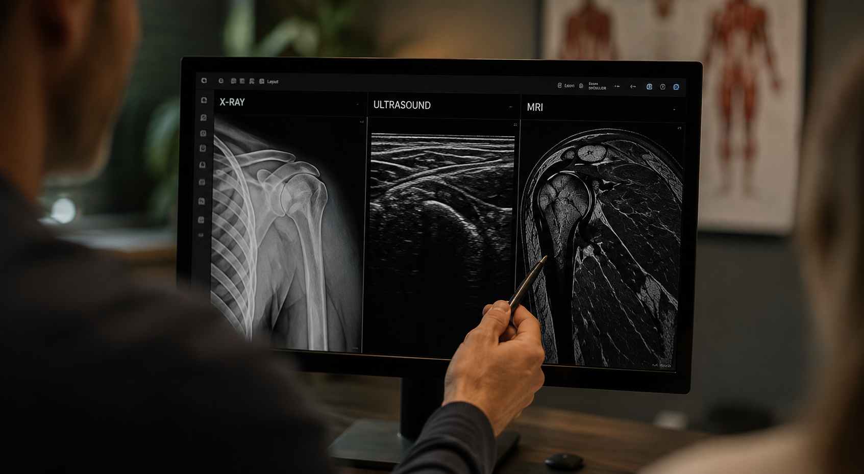
Our typical pathway begins with X-rays, advances to in-office musculoskeletal ultrasound, and then progresses to MRI or other advanced imaging when clinically appropriate.
The goal is not simply to order more imaging.
The goal is to order the right imaging, at the right time, for the right reason—and then use those findings to build a treatment plan.
Our Imaging Pathway
Step 1 — Start With X-Rays
For many orthopedic and musculoskeletal complaints, X-rays are the appropriate first imaging study.
They allow us to evaluate:
Fractures
Joint alignment
Arthritis
Joint-space narrowing
Bone spurs
Calcifications
Degenerative changes
Certain structural abnormalities
Previous surgical hardware
Other bony changes
X-rays are also frequently required by insurance companies before authorization for more advanced imaging such as MRI.
Because of this, beginning with X-rays can often prevent unnecessary delays later in the diagnostic process.
Step 2 — Review the X-Rays + Perform MSK Ultrasound
An X-ray shows us bone exceptionally well.
But many painful orthopedic conditions involve structures that are not well visualized on a standard X-ray.
That is where musculoskeletal ultrasound becomes extremely valuable.
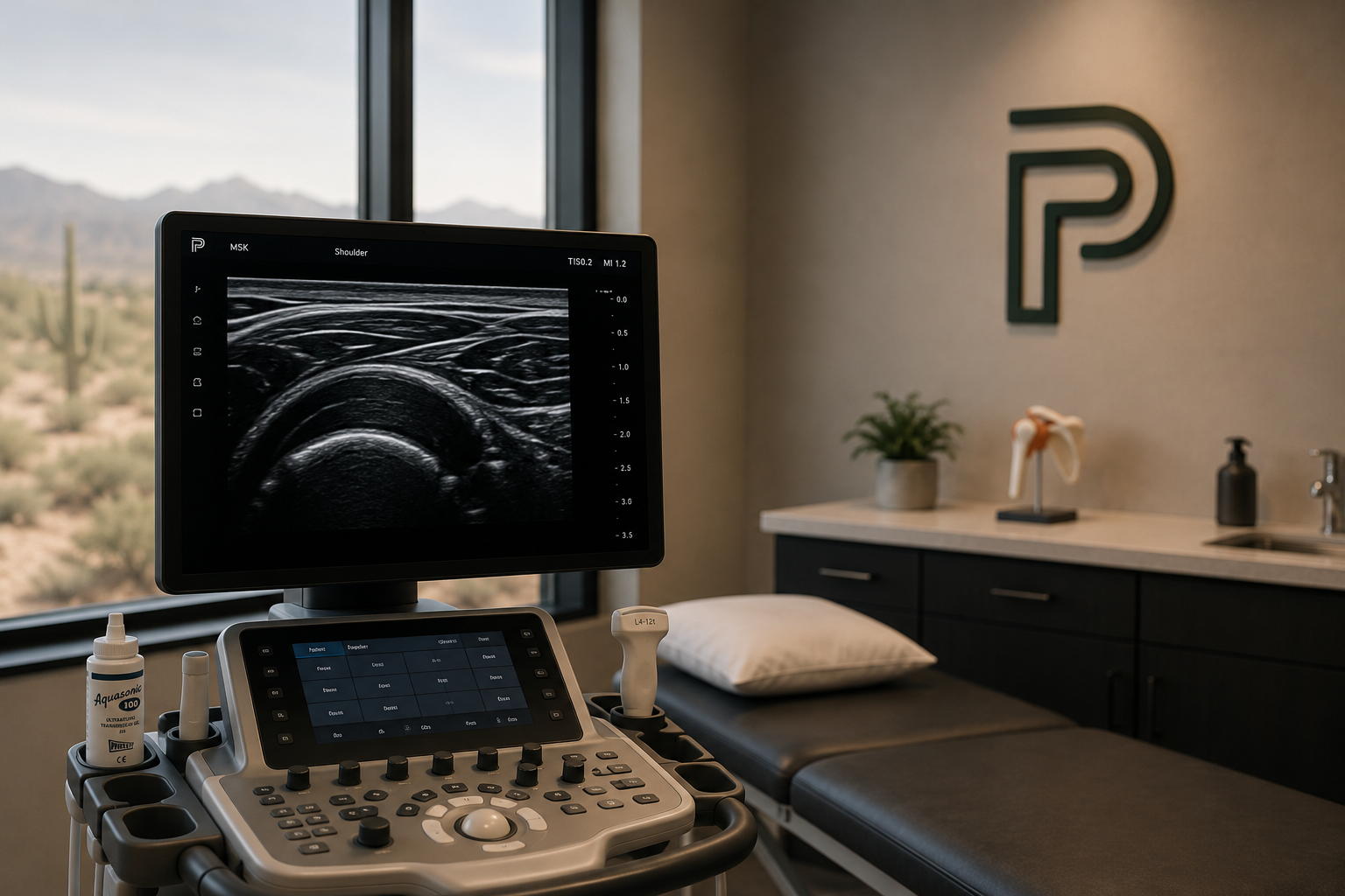
At PRO Sports Medicine, we routinely combine X-ray review with an in-office MSK ultrasound evaluation when appropriate.
Musculoskeletal ultrasound can help us evaluate structures such as:
Tendons
Ligaments
Muscles
Bursae
Joint effusions
Tendon tears
Tendinopathy
Calcifications
Cysts
Soft-tissue abnormalities
Dynamic movement of tendons and joints
Because ultrasound is performed in real time, we can also examine structures while the patient moves or while pressure is applied to the symptomatic area.
This creates an important bridge between the patient's physical examination and the imaging findings.
Two Different Types of Imaging. One Better Clinical Picture.
X-Ray
Best suited for evaluating bone and joint structure.
MSK Ultrasound
Best suited for evaluating many superficial soft tissues dynamically and in real time.
By reviewing these studies together, we often gain a significantly clearer understanding of what may be causing the patient's symptoms.
From there, we determine whether additional imaging is actually necessary.
Step 3 — Determine Whether MRI Is Needed
Not every orthopedic complaint requires an MRI.
After reviewing:
Your symptoms + physical examination + X-rays + MSK ultrasound
we determine whether advanced imaging is likely to change the diagnosis or treatment plan.
When MRI is appropriate, our office can order the study and coordinate the next steps.
MRI may provide additional information regarding:
Rotator cuff injuries
Labral pathology
Meniscus injuries
Ligament tears
Cartilage injury
Muscle injuries
Bone marrow abnormalities
Stress injuries
Disc pathology
Spinal structures
Deeper soft-tissue abnormalities
Complex joint injuries
Conditions not fully characterized on ultrasound or X-ray
The imaging study is selected based on the anatomy and clinical question being investigated.
Why We Don't Automatically Start With MRI
MRI is an extremely valuable diagnostic tool, but more imaging is not always better imaging.
In many situations, the combination of a detailed examination, X-rays, and MSK ultrasound already provides enough information to begin treatment.
Starting with a structured evaluation can:
Reduce unnecessary imaging
Avoid delays caused by insurance authorization
Identify conditions that are visible immediately on ultrasound
Determine whether MRI findings are likely to affect treatment
Create a more efficient path from diagnosis to recovery
Our objective is not to obtain an MRI simply because a patient has pain.
Our objective is to determine whether MRI will provide information that meaningfully affects the next clinical decision.
Step 4 — We Review the MRI With You
Ordering an MRI is only part of the process.
Once the study is completed, we review the results in the context of:
Your symptoms
Your examination
Your X-rays
Your ultrasound findings
Your activity level
Your goals
Previous treatment
The severity and chronicity of the condition
An MRI report alone does not determine treatment.
Many imaging abnormalities can exist without causing symptoms, while some clinically significant injuries may require interpretation within the larger diagnostic picture.
We use the MRI as another piece of evidence—not as the entire diagnosis.
From Imaging to Treatment
Imaging Should Lead Somewhere
The purpose of advanced imaging is to help determine the most appropriate next step.
Depending on the findings, treatment may progress through several levels of care.
Level 1 — Conservative Treatment
For many musculoskeletal conditions, treatment begins conservatively.
This may include:
Physical therapy
Corrective exercise
Activity modification
Home exercise programs
Mobility work
Strengthening
Manual therapy
Recovery strategies
Anti-inflammatory or symptom-management strategies when appropriate
The goal is to restore movement, strength, and function while allowing the injured tissue an opportunity to recover.
Level 2 — Physical Therapy + Biologic Support
When tissue healing, recovery, or chronic injury is a larger component of the problem, we may build a more comprehensive conservative program.
This may include:
Physical therapy + selected peptide therapies
when medically appropriate.
Peptide therapy may be incorporated as one component of a broader recovery strategy rather than replacing rehabilitation.
Movement still matters.
Loading still matters.
Strength still matters.
The objective is to create the best possible biological and mechanical environment for recovery.
Level 3 — Regenerative Medicine & Orthobiologics
When conservative treatment is insufficient—or imaging demonstrates a condition that may benefit from a more advanced nonsurgical approach—we may discuss regenerative medicine.
Depending on the condition, treatment options may include:
Platelet-Rich Plasma (PRP)
PRP + A2M
Plasma-based procedures
Ultrasound-guided injections
Other orthobiologic strategies when clinically appropriate
These treatments are selected based on the specific tissue involved, severity of injury, imaging findings, previous treatment, and patient goals.
Level 4 — Procedural Treatment
Some conditions may require additional procedural intervention.
Depending on the diagnosis, this can include ultrasound-guided procedures or minimally invasive treatment strategies.
For example, chronic tendon pathology may sometimes warrant consideration of procedures such as Tenex, while other conditions may benefit from targeted image-guided interventions.
The treatment pathway is determined by the pathology—not by a predetermined package.
Level 5 — Surgical Referral
Sometimes the correct treatment is surgery.
Our goal is not to keep every patient within conservative or regenerative care indefinitely.
If the examination and imaging indicate that a patient is more likely to benefit from surgical management, we help identify that and facilitate an appropriate referral.
Potential reasons for surgical consultation may include:
Significant tendon rupture
Unstable ligament injury
Advanced structural damage
Severe joint degeneration
Significant mechanical symptoms
Neurologic compromise
Failure of appropriate conservative treatment
Injuries unlikely to respond adequately to nonsurgical care
Knowing when not to continue conservative treatment is just as important as knowing when to offer it.
One Diagnostic Process. Multiple Treatment Pathways.
A major advantage of our approach is continuity.
Instead of obtaining an X-ray at one location, an ultrasound somewhere else, an MRI through another provider, and then trying to determine what to do with all of those results, we help coordinate the diagnostic pathway.
Your pathway may look like:
Clinical Evaluation
↓
X-Ray
↓
X-Ray Review + MSK Ultrasound
↓
Advanced Imaging if Needed
↓
MRI Review
↓
Appropriate Treatment Pathway
Physical Therapy
→ PT + Peptide Therapy
→ Regenerative Medicine / Orthobiologics
→ Procedural Treatment
→ Surgical Referral When Necessary
Not every patient moves through every step.
The pathway becomes more advanced only when the clinical situation requires it.
Why MSK Ultrasound Is Central to Our Approach
Musculoskeletal ultrasound allows us to evaluate many painful structures immediately during the office visit.
That can provide several advantages:
Real-Time Imaging
We can correlate what we see with the exact location of your symptoms.
Dynamic Examination
Tendons, muscles, and joints can be assessed during movement.
Immediate Clinical Integration
The imaging findings can be compared directly with the physical examination.
Image-Guided Treatment
If a procedure is eventually recommended, ultrasound may also be used to precisely guide treatment to the intended structure.
Better MRI Decision-Making
Ultrasound can help clarify which patients are likely to benefit from additional advanced imaging.
We Don't Treat MRI Reports
We Treat Patients.
A radiology report may identify abnormalities, but the presence of an abnormality does not automatically mean it is responsible for your pain.
Likewise, the severity of a patient's symptoms does not always correlate perfectly with the severity of findings described on an imaging report.
Our job is to bring everything together:
History
Physical examination
X-ray
Musculoskeletal ultrasound
MRI when needed
Functional limitations
Treatment response
That complete picture guides our recommendations.
Already Have Imaging?
You do not necessarily need to start over.
If you already have:
X-rays
MRI
CT imaging
Previous ultrasound studies
Radiology reports
bring them with you whenever possible.
We can review previous imaging alongside your current examination and determine whether additional testing is necessary.
In some cases, existing imaging may provide everything we need.
In others, new imaging may be appropriate because the condition has changed or the previous study does not adequately answer the current clinical question.
A More Efficient Route to the Right Treatment
The traditional orthopedic experience can become fragmented.
One provider orders an X-ray.
Another provider orders an MRI.
Another interprets the injury.
Another handles therapy.
And another determines whether an injection or surgery is appropriate.
At Arizona Sports Medicine, we aim to create a more connected process.
Examine.
Image.
Understand.
Treat.
Reassess.
The result is a diagnostic pathway designed around making the next appropriate clinical decision, rather than simply accumulating more tests.
Frequently Asked Questions
Do I need an X-ray before an MRI?
Often, yes. X-rays are commonly the appropriate initial imaging study for musculoskeletal complaints, and many insurance plans also require them before authorizing advanced imaging.
Can you order my MRI?
Yes. When MRI is medically appropriate, we can order advanced imaging based on the suspected condition and clinical question.
Do you perform ultrasound in the office?
Yes. Musculoskeletal ultrasound is an important component of our orthopedic diagnostic process and can often be performed during your evaluation.
Is ultrasound the same as MRI?
No. They provide different types of information. Ultrasound is particularly useful for many superficial soft tissues and dynamic evaluation, while MRI can provide a more comprehensive assessment of deeper structures and certain joint, bone, cartilage, and spinal conditions.
What happens after my MRI?
We review the MRI alongside your examination and previous imaging and then discuss the most appropriate treatment pathway.
Does an abnormal MRI mean I need surgery?
No. Many structural findings can be treated nonsurgically. The significance of an MRI finding depends on symptoms, examination findings, functional limitations, severity, and the specific diagnosis.
What if I have already completed physical therapy?
Previous treatment response is part of the decision-making process. If an appropriate rehabilitation program has failed, advanced treatments may be considered depending on the imaging findings and diagnosis.
What if surgery is the best option?
We will tell you. When surgical evaluation is the appropriate next step, we can recommend referral rather than continuing treatments that are unlikely to solve the underlying problem.
Start With the Right Diagnosis
You should not have to guess whether your pain is coming from a tendon, ligament, joint, muscle, bone, or another structure.
Our diagnostic process is designed to identify the problem systematically and determine how far the workup actually needs to go.
X-Ray. MSK Ultrasound. MRI When Needed.
Then we build the treatment plan around what we find.
Schedule a Musculoskeletal Evaluation
PRO Sports Medicine
Sports Medicine • MSK Ultrasound • Advanced Imaging Coordination • Physical Therapy • Regenerative Medicine • Orthobiologics